Secondary Oral Squamous Cell Carcinoma
Chronic GVH predisposes to the development of other cancers. It is necessary to perform thorough oral examinations to diagnose early oral changes. Documentation (best with photographs) for follow-up or biopsy
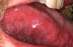
This 45 yr old male patient had an allo HCT 16 years previous for CML. He had chronic GVH of the gingiva but complained of this non-healing ulcer on his tongue for several months. Biopsy proved that it was an early squamous cell carcinoma.

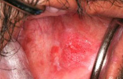
This 42 yr-old male patient had an allo-HCT 3 years previously for AML. He had severe oral GVH and this red pebbly lesion was noted on the left buccal mucosa. It was biopsied and prove to be a squamous cell carcinoma. He developed another squamous cell carcinoma of the lower lip.

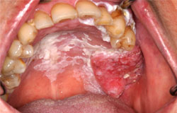
This 70yr-old female had an allo HCT for myelodysplasia in 2001. She had very severe oral, skin and eye GVH resulting in blindness. She presented in 2015 stating that the upper left teeth had spontaneously “fallen out”. Oral examination reveals hyperkeratosis/ veruccous hyperplasia of the hard palate and gingiva of the second quadrant. There is an obvious red and white mass of the left maxillary alveolar ridge extending on to the hard palate. It was biopsied and showed squamous cell carcinoma.

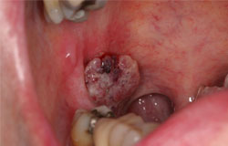
This 73 yr-old male had an allo HCT in 1997 for AML. He had chronic oral GVH and was unaware of this lesion that was noted during a routine dental visit in 2006. An irregular red and white mass emanating from the right tonsil is seen. Biopsy revealed squamous cell carcinoma. He later developed an oral tongue squamous cell carcinoma.

This pantomograph is from a 30yr-old male who had an allo HCT 15 years previously for Fanconi’s anemia. He developed a sore on the posterior right gingiva which was biopsied and proven to be a squamous cell carcinoma. He underwent a partial mandibulectomy but lost the graft and was left with this poor result.

These examples illustrate the potentially long latent period from HCT to the development of the second malignancy. This underscores the importance of a thorough oral examination in all of these patients to detect early oral changes that may require biopsy.