Opportunistic Infections
An increase in the incidence of opportunistic infections such as oral herpetic infections, human papilloma virus infections (e.g. condyloma acuminatum) and candidiasis may also occur. Also, should GVHD develop, medical therapy (systemic) would include immunosuppressant agents. If this occurs, prophylactic antibiotics (as per the current AHA regimen) are advised for dental procedures which will result in gingival bleeding.
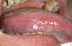
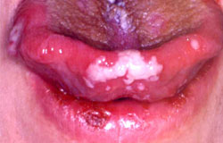
The following two clinical photographs are patients who have had allogeneic bone marrow transplants. They presented with painful oral ulcerations which proved to be positive for Herpes Simplex I and were treated successfully with antiviral agents.


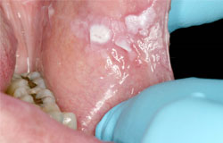
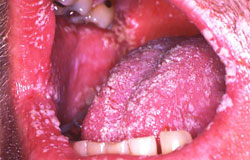
This patient was also post HCT and has numerous white plaques that wiped off revealing erythematous mucosa. Candidiasis was diagnosed and treated with antifungals.

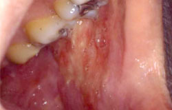
This patient presented post HCT with painful oral ulcers which were CMV positive


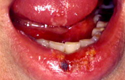
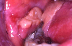
This 57M had a HCT for AML 5 years previous and presented with several recurrent white papules. He had numerous warts on the fingers of both hands as well. Biopsy of the oral lesions were consistent with verucca vulgaris. They recurred for a few years until the lesions on his hands were treated successfully.
Other oral lesions not related to infection or GVH have also been well-documented.
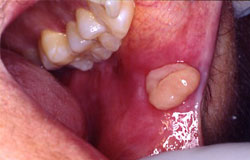
Pyogenic granulomas or hyperplastic granulation tissue have been described post HCT. They are self-limiting and resolve with surgical excision.