Side Effects of Treatment
Side effects of Radiation Therapy to the Head and Neck
Radiation therapy is delivered in daily fractions over several weeks. Most side effects are predictable and expected. Side effects from radiation are usually limited to the area of the patient’s body that is under treatment. One of the aims of modern radiotherapy is to reduce side effects to a minimum, and to help the patient understand and deal with those side effects which are unavoidable.
Acute Side Effects
In the head and neck area, the initial side effects include an increase in saliva viscosity resulting in thick, ropey mucous secretions. Loss of taste or dysguesia is variable and although temporary may last for several months after radiation therapy. Return of normal taste function is less likely in patients over 70 years of age or if the oral tongue is in the volume treated.
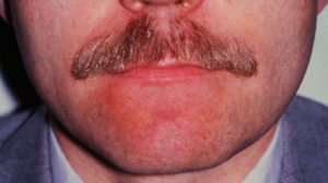
Skin Erythema
Typically the skin starts to become erythematous and sore several days into treatment. The reaction may become more severe during the treatment and for up to about one week following the end of radiotherapy, and the skin may break down. Although this moist desquamation is uncomfortable, recovery is usually quick. Skin reactions tend to be worse in areas where there are natural folds in the skin, such as underneath the female breast, behind the ear, and in the groin.


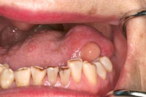
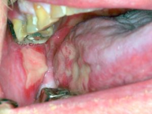
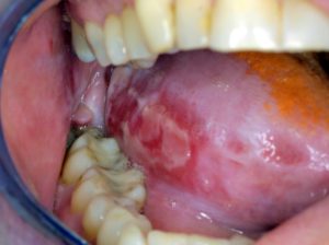
Mucositis
As the epithelium of the oral cavity is turning over rapidly, it is also targeted inadvertently by the radiation resulting in breakdown. Mucositis begins 1-2 weeks into the radiation Confluent ulceration with erythematous borders in a background of generalized “whitening” of the oral mucosa is often seen. This is the major source of pain, dysphagia, weight loss and overall debilitation during radiation therapy. If severe, this can affect swallowing, and the patient may need analgesics and nutritional support/food supplements. A feeding tube may be placed temporarily if the patient has difficulty swallowing. Treatment is empiric and consists of systemic analgesics, topical anaesthetics (mucositis rinse) and change is diet to soft, bland foods.


Fatigue
Fatigue is among the most common symptoms of radiation therapy. Lack of energy, reduced activity and overtired feelings are common symptoms.
Chronic/Long-term effects of Radiation
Dryness
The salivary glands and lacrimal glands have a radiation tolerance of about 30 Gy in 2 Gy fractions, a dose which is exceeded by most radical head and neck cancer treatments. Dry mouth (xerostomia) and dry eyes (xerophthalmia) can become irritating long-term problems and severely reduce the patient’s quality of life. Xerostomia can lead to rampant decay of all remaining teeth over a short time. Dryness is a permanent change and one that many patients have the most difficulty.


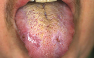

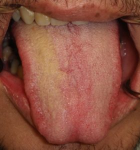
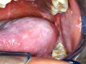
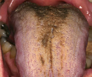
“Hairy tongue”
With a dry mouth, the papillae of the dorsum of the tongue may appear to elongate as bacteria and food debris collect. This may lead to the appearance of a “hairy tongue” that can vary in colour depending on diet and the presence of chromogenic bacteria. Tongue brushing/scraping added to a meticulous oral hygiene regimen can be beneficial.

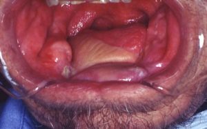
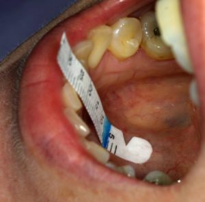
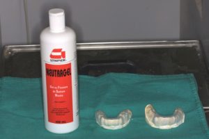
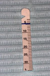
The Modified Schirmer Test (MST) is used in our clinic to measure the approximate salivary flow rate. It is a gross measure of pooled saliva in the floor of mouth measured by the effect of saliva on the Schirmer strips’ capillary action in a calibrated manner as illustrated below. A roughly normal reading is considered to be 30 mm in 2 minutes. Readings are taken prior to radiation therapy, one month post_XRT, 6 months, then annually. The major reason for continuing this measurement after radiation therapy is to reinforce the need for continued Fluoride treatment. Patients will learn to function with a dry mouth and may not feel that it they are as dry subjectively. The measurement is an objective way of indicating that they are still indeed dry and must continue with the fluoride trays daily.


Lymphedema
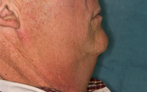
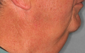
Lymphedema refers to swelling secondary to blockage of lymphatic drainage. With cancer therapy, either radiation therapy or lymph node removal as part of a neck dissection, the lymphatic drainage will be compromised. As there are approximately 300 lymph nodes in the head and neck region, lymphedema in the head and neck region may be significant. The usual signs and symptoms include swelling of the submandibular region giving the appearance of “double chin”. The swelling is related to position and gravity so that it is most noticeable in the morning on awakening and decreases as the day progresses. Sleeping in a more upright position or with a number of pillows will prevent the fluid from flowing in a retrograde fashion. It usually occurs shortly after radiation therapy and is painless. Occasionally there is swelling internally into the tongue and oropharynx. As new drainage pathways develop from the head and neck region to the thoracic duct, lymphedema improves. This is an important side effect that may cause anxiety to the patient who has completed treatment. Massage therapy and exercise are useful in treatment.


Trismus
Tissues which have been irradiated tend to become less elastic over time due to a fibrosis. This can affect the ability to open the mouth (post radiation trismus) making dental care challenging. Trismus may occur after radiation, if the muscles of mastication or other soft tissue components of the oral cavity are in the irradiated volume. It generally does not occur at doses below 30Gy. The normal inter-incisal distance ranges from 35-45 mm which is how trismus treatment is evaluated but solely focusing on this one measurement does not imply success since one can indeed achieve larger inter-incisal distances without helping the patient. Prior to treatment It is critical that other causes of trismus are ruled out (eg infection, pathologic fracture, post-surgical tethering).
Some of the goals of treatment should be to increase the ability of the patient to open their mouth in both the vertical dimension but also to improve the lateral and protrusive movement of the mouth as well as to improve mandibular function. From the patients perspective they want to improve their ability to bite and chew a wide range of foods; speak without disability; have access to maintain oral hygiene and allow dental work; and be able to use their fluoride trays.
Any proposed treatment must avoid damage to the jaws or teeth that can occur when cancer surgery surgical fixation plates are present; individual or multiple teeth have compromised periodontal support; skin or mucosa of the oral aperture is susceptible to tearing; and mandibular bone is weakened by virtue of its being irradiated, surgically altered or otherwise compromised (for example by having an impacted third molar present).
Prior to commencement these structures must be examined and their health documented.
There are two philosophies for treatment. Passive mouth-movement exercise involve the patient doing their own mouth opening and mouth-moving exercises regularly during and after radiation in order to maintain as much jaw mobility as possible. This is likely not effective after trismus is already present and may be problematic for patients with oral mucositis.
Other possibilities for management include devices that open the jaws by using mechanical leverage. Two devices are currently available Orastretch and Therabite (we may need permission to photo these unless we buy them)
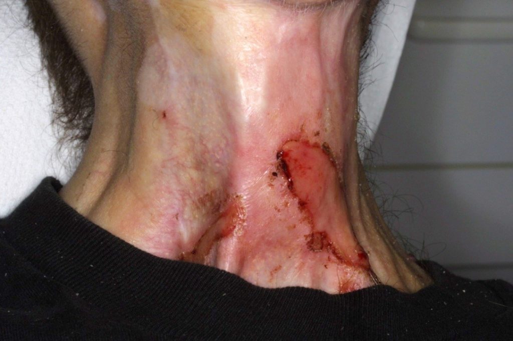
Radiation-induced tumors
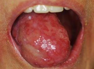
A well-documented late effect of radiation therapy involves the development of secondary new malignancies at the site of previous radiation. This effect is rare, occurring in 0.1% and can occur 5 – 30 years after initial therapy. The new tumour occurs within the field of radiation and are usually sarcomas. In bone, the most common type is osteosarcoma and when affecting soft tissues, undifferentiated pleomorphic sarcoma (formerly malignant fibrous histiocytoma) are the histologic types. The significance of post radiation sarcomas is the poor prognosis. This is usually related to factors such as delay in diagnosis leading to large unresectable lesions, older age, anaplastic nature and lack of effective adjuvant treatment.
This 74 yr-old female was treated for a primary tongue carcinoma with surgery and post-operative radiation therapy in 2000. She developed this ulcerated mass 11 years after her primary therapy. This was diagnosed as a spindle cell (sarcomatoid) carcinoma and was treated surgically.