Malignant Hematology
Bone Marrow/Stem cell Transplants
The Dentists Role
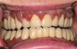
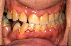
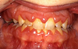
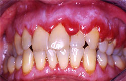
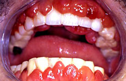
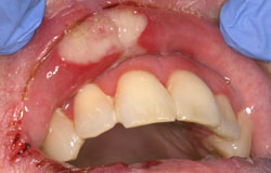
It is well documented that oral manifestations of systemic disease may be the presenting chief complaint. The dentist must be aware of these and act appropriately to aid in the diagnosis. For example, acute myelogenous leukemia (AML) may present with gingival involvement. In our experience of screening leukemia patients, this occurs in less than 2% of the newly diagnosed leukemics. However, it is a useful sign of underlying disease that may present to the dentist first. One of the distinguishing features from generalized hyperplastic gingivitis is the presence of gingival pain and hemorrhage when there are leukemic infiltrates in the gingiva. The following are examples of varying degrees of involvement of the gingiva in patients diagnosed with AML. With successful treatment of the leukemia, the gingival hyperplasia regresses. Until that time, empiric treatment with chlorhexidine mouthwashes is indicated.


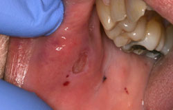
Another common finding may be petechia or ecchymosis with minor trauma secondary to thrombocytopenia as seen in the following example.

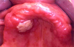
Neutropenic ulcers may occur when a traumatic ulcer becomes secondarily infected because of the lack of immunoprotection of the neutrophils. These painful ulcers appear large and covered with a thick coating resembling bacterial cultures. Often the oncologist will refer the patient for biopsy of these lesions to rule out leukemic infiltrates. Caution must be exercised as the patient is often thrombocytopenic as well as neutropenic and excessive bleeding may result. The following 2 photographs are examples of neutropenic ulcerations.

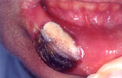

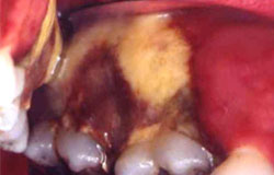
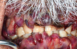
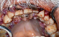
This 66 yr-old male who was being treated with chemotherapy (Azacytadine) for myelodysplasia was referred for assessment and biopsy of this ulcer. He stated that he developed right facial swelling 4 days previously and felt that he had bitten his cheek during the night. His neutrophil count was 0.17 and his platelet count was 7. There was a 2 cm in diameter ulcer on a raised violaceous base. The ulcer had a black covering with a purulent exudate. Given the history of trauma and his blood counts, the impression of an infected neutropenic ulcer was entertained. He was placed on antibiotics and chlorhexidine mouth rinse and the following photograph was taken one week later.

Although the blood counts remained low (neutrophil count 0.20), the ulcer and swelling have improved significantly in one week. The swelling was presumed to be a hematoma from the trauma and thrombocytopenia. Note the petechiae on the buccal mucosa indicative of thrombocytopenia.

The following example is a 59 yr-old male who received an allogeneic bone marrow transplant for AML 2 months previously. He developed several large painful ulcers of which this was an example. His neutrophil count was 11 and the oncololgist referred him for biopsy of an ulcer to rule out graft vs. host disease. He had a previous episode prior to transplant of large oral ulcerations that resolved over a 6 week period. A biopsy was performed and the pathologic report was “non-specific ulcer”. This is consistent with aphthous major and given the previous history, the most likely diagnosis. He was being treated for GVH of the GI and his ulcers healed presumably with this treatment.

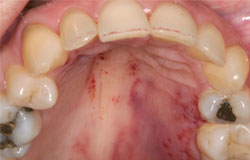
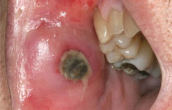
The following example shows another ulcer in a patient with leukemia. The patient is an 85 yr-old male who was on chemotherapy for AML but not in remission. He developed this ulcer on the palate such that he had difficulty wearing his complete upper denture. The ulceration appears secondarily infected as well. The ulcer was biopsied and revealed a leukemic infiltrate.

The previous examples illustrate that ulcerations in patients with leukemia or post-HCT may be difficult to diagnose. The differential diagnosis should include neutropenic ulcer, leukemic infiltrate, aphthous, chemotherapy related mucositis and infection. Careful review of the history and blood counts as well as documentation of the lesion are necessary to determine the best course of treatment for the patient. For example, topical steroids would be contraindicated for a viral ulcer. Oral pathology consultation if available is optimal.