Other non GVH Oral Lesions
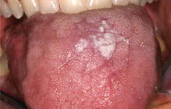
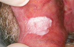
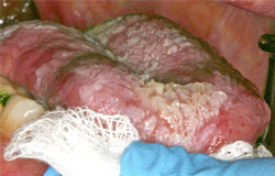
Verrucous hyperplasia
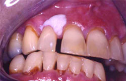
Isolated well-demarcated white patches and plaques are also encountered post HCT. Histologically, they are described as hyperkeratosis with a veruccous surface morphology. Usually they are asymptomatic and require no treatment other than close follow-up.


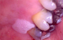

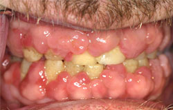
This last example also shows extensive root caries secondary to dry mouth.
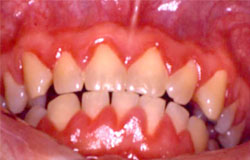
Drug-induced gingival hyperplasia is well-documented with phenytoin, cyclosporine, diltiazem, amlodopine and others. As cyclosporine is an immunosuppressant that is often used in the treatment of GVH, it is important to distinguish it from leukemic gingivitis. The first example below is a patient post HCT with cyclosporine induced gingival hyperplasia. Biopsy was necessary to rule out relapsed AML. The second example is a more classic picture of the fibrotic gingival overgrowth seen with cyclosporine. Once the diagnosis is confirmed, communication with the oncologist may prompt the use of an alternative immunosuppressant.